38 human heart diagram with labels
Human Heart Diagram Teaching Resources | Teachers Pay Teachers Human Heart Parts and Blood Flow Labeling Worksheets - Diagram/Graphic Organizer. by. TechCheck Lessons. 21. $2.25. Zip. This resource contains 2 worksheets for students to (1) label the parts of the human heart and (2) Fill in a flowchart tracing the path of blood flowing though the circulatory system. Answer keys included. Diagrams, quizzes and worksheets of the heart | Kenhub Worksheet showing unlabelled heart diagrams. Using our unlabeled heart diagrams, you can challenge yourself to identify the individual parts of the heart as indicated by the arrows and fill-in-the-blank spaces. This exercise will help you to identify your weak spots, so you'll know which heart structures you need to spend more time studying ...
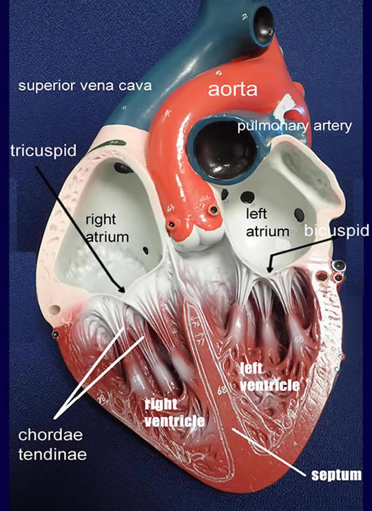
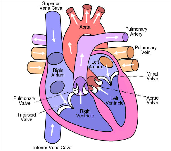
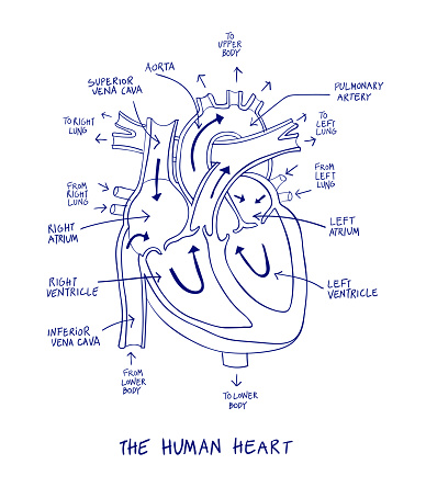
Label Heart Anatomy Diagram Printout - EnchantedLearning.com Label Heart Interior Anatomy Diagram: Human Anatomy: The heart is a fist-sized, muscular organ that pumps blood through the body. Oxygen-poor blood enters the right atrium of the heart (via veins called the inferior vena cava and the superior vena cava). The blood is then pumped into the right ventricle and then through the pulmonary artery to ...

Human heart diagram with labels
Human Heart Diagram Labeled | Science Trends Human Heart Diagram Labeled Daniel Nelson 1, January 2019 | Last Updated: 3, March 2020 The human heart is an organ responsible for pumping blood through the body, moving the blood (which carries valuable oxygen) to all the tissues in the body. Without the heart, the tissues couldn't get the oxygen they need and would die. Human Heart: Label the diagram 1 - Liveworksheets Human Heart: Label the diagram 1 Study the figure carefully.Label the 10 parts of the human heart A-J. ID: 1781041 ... Age: 14+ Main content: Human Circulatory System Other contents: Human Heart Add to my workbooks (15) Download file pdf Embed in my website or blog Add to Google Classroom Add to Microsoft Teams Share through Whatsapp: Human Heart - Diagram and Anatomy of the Heart - Innerbody The heart is a muscular organ about the size of a closed fist that functions as the body's circulatory pump. It takes in deoxygenated blood through the veins and delivers it to the lungs for oxygenation before pumping it into the various arteries (which provide oxygen and nutrients to body tissues by transporting the blood throughout the body).
Human heart diagram with labels. A Diagram of the Heart and Its Functioning Explained in Detail The heart blood flow diagram (flowchart) given below will help you to understand the pathway of blood through the heart.Initial five points denotes impure or deoxygenated blood and the last five points denotes pure or oxygenated blood. 1.Different Parts of the Body ↓ 2.Major Veins ↓ 3.Right Atrium ↓ 4.Right Ventricle ↓ 5.Pulmonary Artery ↓ 6.Lungs Label the Heart Diagram | Quizlet Start studying Label the Heart. Learn vocabulary, terms, and more with flashcards, games, and other study tools. Human Heart Diagram - Side View and Top View As shown below, this human heart diagram clearly illustrates the valves of the heart. The valves illustrated below are the pulmonary, tricuspid, aortic and mitral valve. So you know, I had the aortic and pulmonary valves of my heart replaced via the Ross Procedure. Heart Diagram with Labels and Detailed Explanation - BYJUS The human heart is the most crucial organ of the human body. It pumps blood from the heart to different parts of the body and back to the heart. The most common heart attack symptoms or warning signs are chest pain, breathlessness, nausea, sweating etc. The diagram of heart is beneficial for Class 10 and 12 and is frequently asked in the ...
Human Heart (Anatomy): Diagram, Function, Chambers, Location in Body The heart is a muscular organ about the size of a fist, located just behind and slightly left of the breastbone. The heart pumps blood through the network of arteries and veins called the... A Labeled Diagram of the Human Heart You Really Need to See The human heart, comprises four chambers: right atrium, left atrium, right ventricle and left ventricle. The two upper chambers are called the left and the right atria, and the two lower chambers are known as the left and the right ventricles. The two atria and ventricles are separated from each other by a muscle wall called 'septum'. Human Heart - Diagram and Anatomy of the Heart - Innerbody The heart is a muscular organ about the size of a closed fist that functions as the body's circulatory pump. It takes in deoxygenated blood through the veins and delivers it to the lungs for oxygenation before pumping it into the various arteries (which provide oxygen and nutrients to body tissues by transporting the blood throughout the body). Human Heart: Label the diagram 1 - Liveworksheets Human Heart: Label the diagram 1 Study the figure carefully.Label the 10 parts of the human heart A-J. ID: 1781041 ... Age: 14+ Main content: Human Circulatory System Other contents: Human Heart Add to my workbooks (15) Download file pdf Embed in my website or blog Add to Google Classroom Add to Microsoft Teams Share through Whatsapp:
Human Heart Diagram Labeled | Science Trends Human Heart Diagram Labeled Daniel Nelson 1, January 2019 | Last Updated: 3, March 2020 The human heart is an organ responsible for pumping blood through the body, moving the blood (which carries valuable oxygen) to all the tissues in the body. Without the heart, the tissues couldn't get the oxygen they need and would die.


![BIOMED ALL INVITED: The Human Heart [ANATOMY/PHYSIOLOGY/CONDUCTION SYSTEM]](https://blogger.googleusercontent.com/img/b/R29vZ2xl/AVvXsEhVPNgp2meK6qlRuRU7FaZEh3K0LNV5gYTJWYWTh9nWubRlhXFX1LCsUjBC5RoXtRAEIoW_vnCyad9iVGzLuzSQL0Cyl8X7ZmE7nM0FaXL5gX-W8cqha_CPOSrvjdj6OVk7zJd4gddBym0f/s1600/heart+anatomy.jpg)


Post a Comment for "38 human heart diagram with labels"