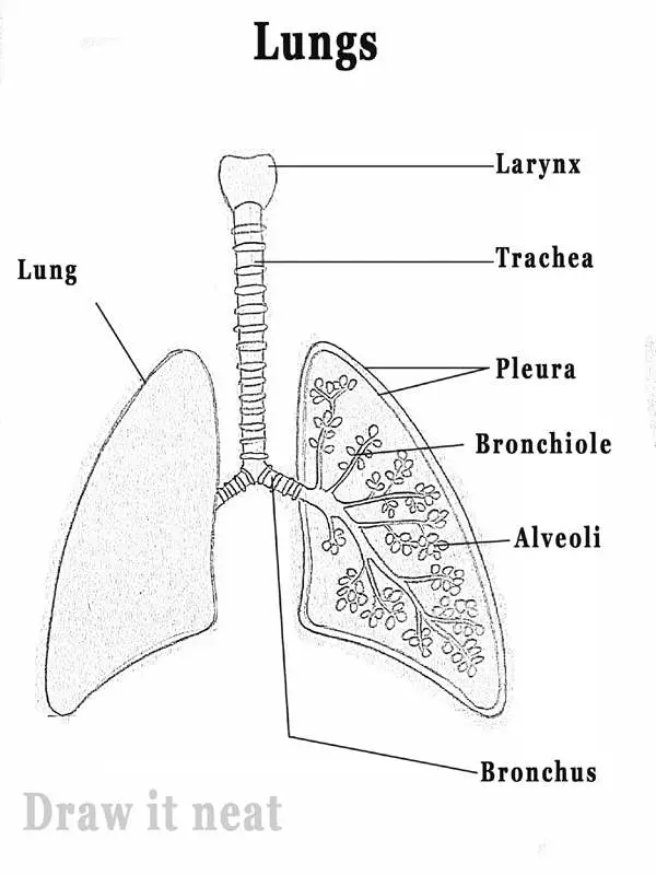
41 diagram of the lungs with labels
Diagram Of The Respiratory System With Labels Stock Photos, Pictures ... Browse 154 diagram of the respiratory system with labels stock photos and images available, or start a new search to explore more stock photos and images. Newest results The respiratory system Lungs with Alveoli Labeled The digestive system lung. Human Respiratory System Lungs Label Design Anatomy Human Lungs Human Lungs Diagram Nervous System Worksheet Answers - WikiEducator Jan 14, 2008 · 8. The diagram below shows a section of a dog’s brain. Add the labels in the list below and, if you like, colour in the diagram as suggested. Cerebellum - blue; Spinal cord - green; Medulla oblongata - orange; Hypothalamus - purple; Pituitary gland - red; Cerebral hemispheres – yellow. 9. Match the descriptions below with the terms in the list.
Label the lung diagram - Quizlet Start studying Label the lung diagram. Learn vocabulary, terms, and more with flashcards, games, and other study tools.

Diagram of the lungs with labels
Digram Of The Lungs - Agaliprogram Diagram of the lungs with labels labeling of the lungs label the lungs diagram diagram of lungs with. By admin apr 15, 2019. Source: breathmatters.com. Lungs are the largest organ in the respiratory system. The lungs are roughly cone shaped with an apex base three surfaces and three borders. Diagram Lungs Stock Illustrations - 2,523 Diagram Lungs ... - Dreamstime Download 2,523 Diagram Lungs Stock Illustrations, Vectors & Clipart for FREE or amazingly low rates! New users enjoy 60% OFF. 187,560,892 stock photos online. ... Labeled diagram with brain sections. Cranial nerves vector illustration. Labeled diagram with brain sections and its. Lungs. Respiratory organs detailed anatomy illustration on a ... Parents (for Parents) - Nemours KidsHealth They still put nicotine or chemicals in the body and can damage the lungs. Get the facts. Managing Your Toddler's Behavior. Learn how to encourage good behavior, handle tantrums, and keep your cool when parenting your toddler. Questions and Answers. How can I teach my kids to be smart on social media?
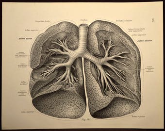
Diagram of the lungs with labels. Lung Diagram Labeled | EdrawMax Template in the following lung labeled diagram, we have shown thyroid cartilage, cricoid cartilage, tracheal cartilage, apex, left upper lobe, hilum, left bronchus, oblique fissure, bronchioles, left lower lobe, base of lung, cardiac notch, right lower lobe, oblique fissure, right middle lobe, horizontal fissure, right bronchus, right upper lobe, and … The Respiratory System (Label Diagram) - ScienceQuiz.net Match each pair by dragging from right to left. When complete click Check button. Diagram Of Lungs Below is a blank diagram, followed by the labeled diagram with the answers. To help adjust your breathing to changing needs, your body has sensors that send signals to the breathing centers in the brain. It leads to a bluish coloration of skin due to lack of oxygen , shortness of breath, and chest pain among other symptoms. › enAnatomy, medical imaging and e-learning for ... - IMAIOS IMAIOS and selected third parties, use cookies or similar technologies, in particular for audience measurement. Cookies allow us to analyze and store information such as the characteristics of your device as well as certain personal data (e.g., IP addresses, navigation, usage or geolocation data, unique identifiers).
› circulatory-system-diagramCirculatory System Diagram - SmartDraw They may come with or without labels. Common circulatory system diagrams show pulmonary circulation, coronary circulation, systematic circulation, veins, arteries, or a combination. The systemic circulation system is the most commonly illustrated of the systems that make up the circulatory system as it is the largest. Anatomy, medical imaging and e-learning for healthcare IMAIOS and selected third parties, use cookies or similar technologies, in particular for audience measurement. Cookies allow us to analyze and store information such as the characteristics of your device as well as certain personal data (e.g., IP addresses, navigation, usage or geolocation data, unique identifiers). Label Lungs Diagram Printout - EnchantedLearning.com Read the definitions below, then label the lung anatomy diagram. bronchial tree - the system of airways within the lungs, which bring air from the trachea to the lung's tiny air sacs (alveoli). cardiac notch - the indentation in the left lung that provides room for the heart. diaphragm - a muscular membrane under the lungs. Label the heart — Science Learning Hub Jun 16, 2017 · Drag and drop the text labels onto the boxes next to the diagram. Selecting or hovering over a box will highlight each area in the diagram. In this interactive, you can label parts of the human heart. ... Receives oxygenated blood from the lungs. Left ventricle. Region of the heart that pumps oxygenated blood to the body. Pulmonary artery ...
Diagram Of The Lung Diagram of the lungs 652. Width: 600, Height: 600, Filetype: jpg Create healthcare diagrams like this example called lobes of the lung in minutes with smartdraw. Width: 975, Height: 731, Filetype: jpg Lungs diagram below displays human lungs anatomy consisting of bronchi lobes alveoli diaphragm bronchioles pleura trachea. Circulatory System Diagram - Cardiovascular System and Blood ... They may come with or without labels. Common circulatory system diagrams show pulmonary circulation, coronary circulation, systematic circulation, veins, arteries, or a combination. The systemic circulation system is the most commonly illustrated of the systems that make up the circulatory system as it is the largest. Diagram Of The Lungs With Labels Labeling Of The Lungs Label The Lungs ... Diagram Of The Lungs With Labels Labeling Of The Lungs Label The Lungs Diagram Diagram Of Lungs With. By admin Apr 15, 2019. Share this page . Post navigation. Lung Lobectomy: What you need to know . By admin. Related Post. Leave a Reply Cancel reply. You must be logged in to post a comment. Labeled diagram of the lungs/respiratory system. - SERC View Original Image at Full Size. Labeled diagram of the lungs/respiratory system. Image 37789 is a 1125 by 1408 pixel PNG Uploaded: Jan10 14. Last Modified: 2014-01-10 12:15:34
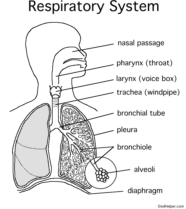
› photos › human-throat-anatomyHuman Throat Anatomy Pictures, Images and Stock Photos Human Respiratory System anatomical vector illustration, medical education cross section diagram with nasal cavity, throat, lungs and alveoli. Human Respiratory System anatomical vector illustration, medical education cross section diagram with nasal cavity, throat, esophagus, trachea, lungs and alveoli. human throat anatomy stock illustrations
Label The Organs Of The Body Teaching Resources | TpT Human Body Organ PostersThis resource includes 26 colored and black and white posters of the organs, include the bladder, brain, eyeball, gallbladder, heart, kidneys, large intestine, small intestine, liver, lungs, pancreas, skin, and stomach. Each poster includes a label, illustration, and description.
Lobes of the Lung - SmartDraw Lobes of the Lung. Create healthcare diagrams like this example called Lobes of the Lung in minutes with SmartDraw. SmartDraw includes 1000s of professional healthcare and anatomy chart templates that you can modify and make your own. 4/22 EXAMPLES. EDIT THIS EXAMPLE.
Lungs (Human Anatomy): Picture, Function, Definition, Conditions - WebMD Lung Tests. Chest X-ray: An X-ray is the most common first test for lung problems.It can identify air or fluid in the chest, fluid in the lung, pneumonia, masses, foreign bodies, and other ...
Lungs Diagram Labeled Pictures, Images and Stock Photos Search from Lungs Diagram Labeled stock photos, pictures and royalty-free images from iStock. Find high-quality stock photos that you won't find anywhere else.
PDF ANATOMY OF LUNGS - University of Kentucky SURFACES OF THE LUNG 1. Costal Surface- It is in contact with costal pleura and overlying thoracic wall. 2. Medial Surface- Posterior / Vertebral Part - Anterior / Mediastinal Part Relations of Posterior Part 1. Vertebral Part 2. Intervertebral Discs 3. Posterior Intercostal Vessels 4. Splanchic Nerves RELATIONS OF ANTERIOR PART RIGHT SIDE 1.
Lung Diagram | Free Lung Diagram Template - Edrawsoft The lung diagram template here clearly presents a pair of spongy on both side of the chest. Simply hitting on the template to learn more parts including pleura, ribs, bronchi, alveoli and more. Feel free to find out more human anatomy templates and symbols in the free download version.
A Guide to Understand Lung with Diagrams | EdrawMax Online Source: EdrawMax Online. Step 2: The students can make several types of science diagrams on this tool. To get the lung diagram, they need to go to the human anatomy option. They can find the lung diagram in this tab. Source: EdrawMax Online. Step 3: They can take the image and then edit it out as per their choice.
Label the Lungs Diagram | Quizlet Start studying Label the Lungs. Learn vocabulary, terms, and more with flashcards, games, and other study tools.
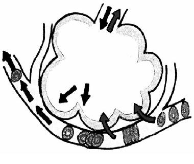
Fully Labelled Diagram Alveolus Lungs Showing Stock ... - Shutterstock Shutterstock customers love this asset! Stock Vector ID: 369984683 Fully labelled diagram of the alveolus in the lungs showing gaseous exchange. Vector Formats EPS 1114 × 800 pixels • 3.7 × 2.7 in • DPI 300 • JPG Vector Contributor S Steve Cymro Similar images Assets from the same collection Similar video clips
Lungs label - Teaching resources Lungs - The Lungs - Lungs Diagram - AC 4.1. Question 17 - Label the main components of the human lungs - The Lungs - The Lungs - Structure of the Lungs . Community ... Lung Label Labelled diagram. by Selina13. Haythorne German label hobby verbs Labelled diagram. by Haythorneg. KS3 German. Label a plant Y1 Labelled diagram. by Helen123.
Female Anatomy Diagram High Resolution Stock Photography … Find the perfect female anatomy diagram stock photo. Huge collection, amazing choice, 100+ million high quality, affordable RF and RM images. ... Woman holding a blackboard with a titled diagram / illustration of human Lungs drawn chalk. ... with labels. Map on Skin of Cutaneous Sensory Innervation of ...
› photos › muscular-systemMuscular System Labeled Diagram Stock Photos, Pictures ... Lungs and bronchi disease with breathing problems diagram. Asthmatic and normal airway cross section with labeled structure and symptoms. muscular system labeled diagram stock illustrations Asthma vector illustration.
› labelling_interactives › 1Label the heart — Science Learning Hub Jun 16, 2017 · Labels. Description. Vena cava. Carries deoxygenated blood from the body to the heart. Semilunar valve. Flaps that prevent backflow of blood. Left atrium. Receives oxygenated blood from the lungs. Left ventricle. Region of the heart that pumps oxygenated blood to the body. Pulmonary artery. Carries deoxygenated blood to the lungs. Right ventricle
Heart Diagram with Labels and Detailed Explanation - BYJUS The diagram of heart is beneficial for Class 10 and 12 and is frequently asked in the examinations. A detailed explanation of the heart along with a well-labelled diagram is given for reference. ... The pulmonary artery, being an exception, carries deoxygenated blood to the lungs for purification. The veins carry impure blood from different ...
Lung Diagram Labelling Activity | Primary Resources | Twinkl This handy Lung Labelling Worksheet gives your children the opportunity to show how much they've learned about the human lung system. The beautifully hand-drawn illustration shows a lung diagram, labelled with blank spaces where learners can fill in its different components. Encourage your students to work independently and label the parts of the lungs they can see. This teaching resource also ...
Muscular System Labeled Diagram Pictures, Images and Stock … Frontal view of the muscular system of the male human body with descriptive labels pointing to the muscles on a white background. ... Disease with breathing problems diagram.. Asthma vector illustration. Lungs and bronchi disease with breathing problems diagram. Asthmatic and normal airway cross section with labeled structure and symptoms ...
Labeled Diagram of the Human Lungs - Bodytomy Given below is a labeled diagram of the human lungs followed by a brief account of the different parts of the lungs and their functions. Each lung is enclosed inside a sac called pleura, which is a double-membrane structure formed by a smooth membrane called serous membrane.




Post a Comment for "41 diagram of the lungs with labels"