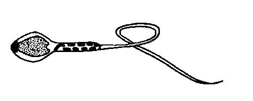
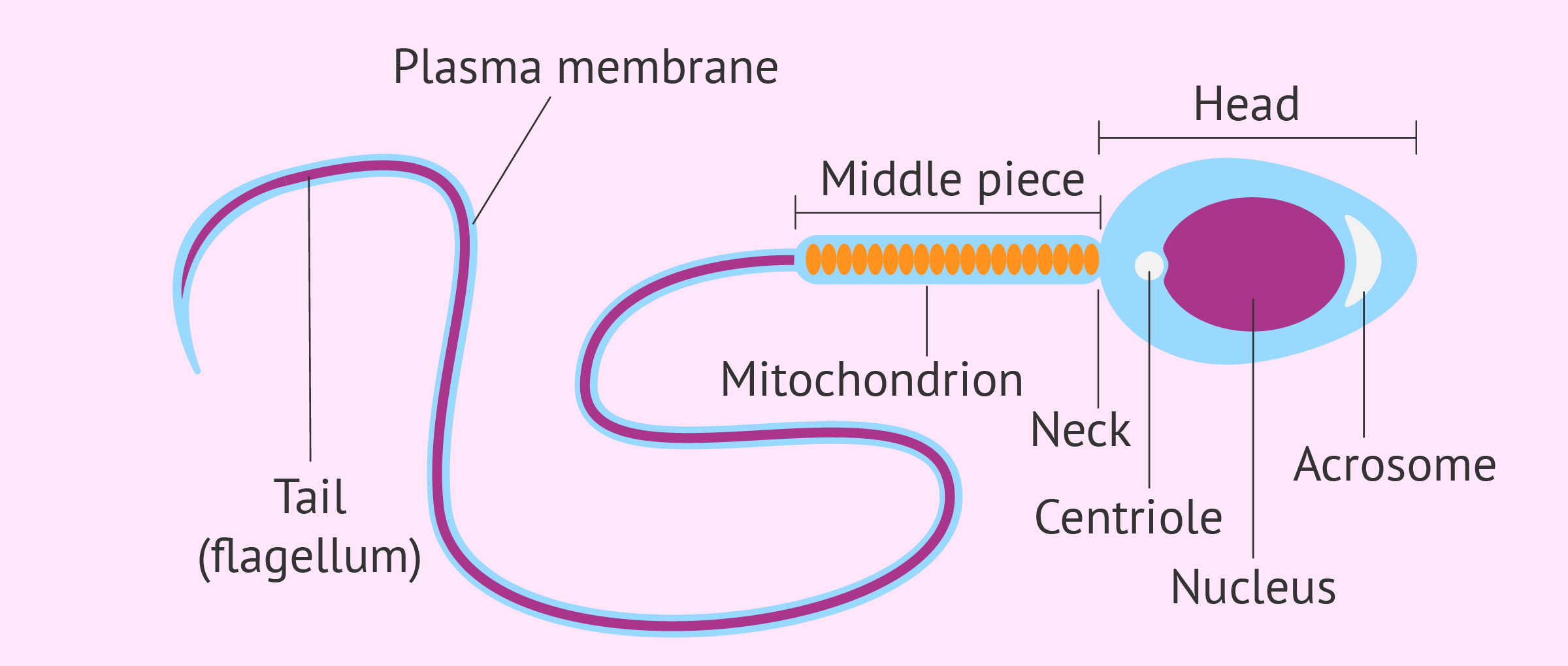
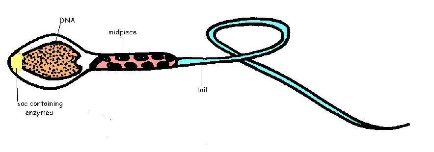
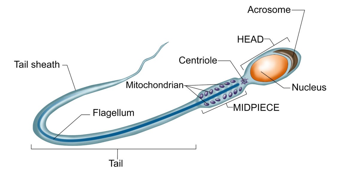
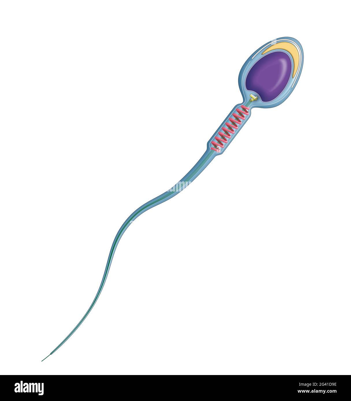
44 sperm cell diagram with labels
Sperm Cells Images | Free Vectors, Stock Photos & PSD Find & Download Free Graphic Resources for Sperm Cells. 700+ Vectors, Stock Photos & PSD files. Free for commercial use High Quality Images Sperm Cell, Egg Cell Diagram Label Worksheets (Differentiated) Three excellently differentiated worksheets. Engaging activity where pupils have to label the different parts of the male and femal gametes. Very well structured and scaffolded according to ability (from SEN to high ability). Excellent for visual learners. Compatible with all biology exam boards (including AQA, Edexcel, OCR).
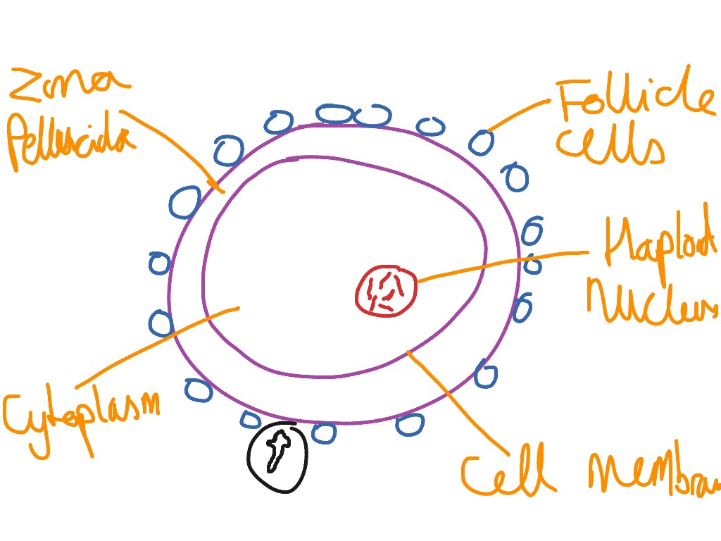
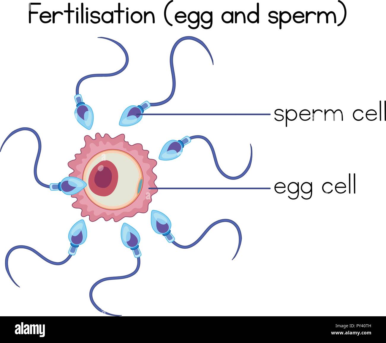
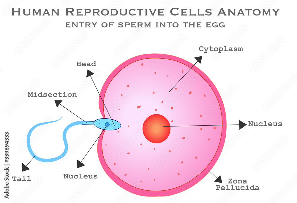
Specialised animal cells - Cell structure - Edexcel - GCSE Biology ... The tail enables the sperm to swim. Sperm are the smallest cells in the body and millions of them are made. Egg cell. The cytoplasm contains nutrients for the growth of the early embryo. The ...
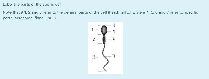
Sperm cell diagram with labels
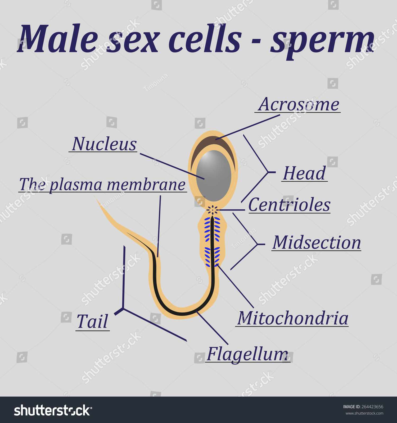
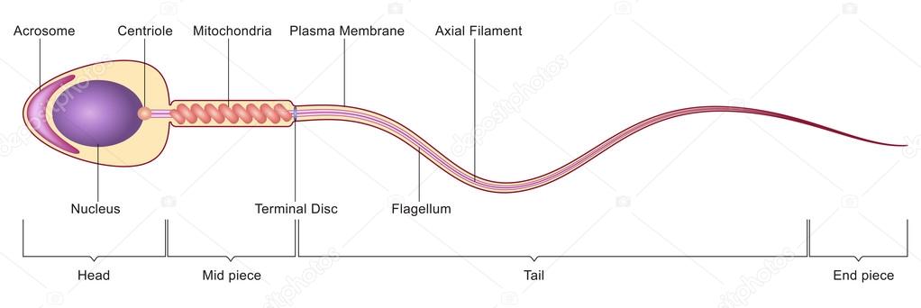
The above diagram is of the sperm cell. - Toppr Ask The above diagram is of the sperm cell. (a) Acrosome: It contains enzymes used for penetrating the female egg. ... Draw the diagram of human sperm and label its parts. Write few lines about it. Medium. View solution > View more. More From Chapter. Human Reproduction. View chapter > Revise with Concepts. Advanced Knowledge of Fertilization in ... Sperm Diagram Stock Photos, Pictures & Royalty-Free Images - iStock structure of a sperm cell Human Sperm cell Anatomy Penis or Male Reproductive System is a 3D illustration. Comparison between normal and low sperm count Spermatozoons Group Flowing Toward Female Ovum Egg Group of Spermatozoons Flowing Toward Female Ovum Egg Isolated on White Background. Natural Fertilization Process Microscopic View. Sperm Under Microscope with Labeled Diagram - AnatomyLearner Sperm Under Microscope with Labeled Diagram 24/06/2022 17/06/2022 by anatomylearner While studying the histological features of the seminiferous tubules and epididymis, you will see sperm cells under the microscope. They are much smaller and lie in groups along the inner margin of the Sertoli cells.
Sperm cell diagram with labels. Structure of Human Sperm: Check Types of Sperm - Embibe - Embibe Exams Explain the Structure of Human Sperm with Labelled Diagram Fig: Structure of a sperm cell Learn Exam Concepts on Embibe What is the Structure of Sperm? Human sperm is a microscopic structure whose shape is like a tadpole. It has flagella which make it motile. Its diameter is \ (2 - 5 {\rm { \mu m}},\) and its length is \ (60 {\rm { \mu m}}.\) Testes: Anatomy and Function, Diagram, Conditions, and Health Tips The epididymis stores sperm cells until they're mature and ready for ejaculation. ... Explore the interactive 3-D diagram below to learn more about the testes. ... (2015). "Off-label" usage ... Draw the diagram of human sperm and label its parts. Write few lines ... Draw the diagram of human sperm and label its parts. Write few lines about it. Medium Solution Verified by Toppr The sperm cells are the haploid gametes which are produced in the male. There are different parts of the sperm cell. (a) Acrosome: This structure contains enzymes used for penetrating the female egg. Structure Of Sperm Diagram || Draw Labelled Diagram Of Sperm - YouTube Hello Everyone.Structure Of Sperm Diagram || Draw Labelled Diagram Of Sperm || Class 12 || BiologyStructure Of Sperm Diagram, Draw Labelled Diagram Of Sperm,...
Spermatogenesis Diagram & Function | What is the Process of Sperm ... Beneath the Sertoli cells are the spermatogonia, which are germ cells that will go through mitosis and ultimately create sperm. In humans, each day, roughly 25 million spermatogonia divide, and... Testes - Anatomy Pictures and Information - Innerbody The testes (singular: testis), commonly known as the testicles, are a pair of ovoid glandular organs that are central to the function of the male reproductive system. The testes are responsible for the production of sperm cells and the male sex hormone testosterone. How to draw Sperm Cell || Study of Human Spermatozoon diagram and label ... 'How to draw Sperm Cell || Study of Human Spermatozoon diagram and label the parts' is demonstrated in this video tutorial step by step.Sperm is the male rep... Sperm Cell - The Definitive Guide | Biology Dictionary A sperm cell or spermatozoon is a gamete (sex cell) produced in the male reproductive tract. It is a motile cell with a single aim - to fertilize a female egg. Each sperm cell contains the entire genome of the male that produces it. In combination with the female genome contained within the egg, a zygote is formed - a single totipotent stem ...
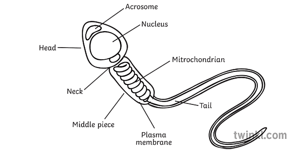
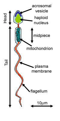
Sperm Cells - Definition, Function, Structure, Adaptations, Microscopy The head of the sperm measures 2.5 to 3.5 um in diameter and 4.0 to 5.5 um in length (um=micrometers). This results in a 1.50 to 1.70 length to width ratio They have a well-developed acrosome that covers 40 to 70 percent of the oval shaped head A slim middle section (body) that is approximately the same length as the head Sperm Cell Labelled Illustration - Twinkl Sperm Cell Labelled. Use this image now, for FREE! Create your own Sperm Cell Labelled themed poster, display banner, bunting, display lettering, labels, Tolsby frame, story board, colouring sheet, card, bookmark, wordmat and many other classroom essentials in Twinkl Create using this, and thousands of other handcrafted illustrations. Diagram and label sperm cell Diagram | Quizlet Terms in this set (4) Midsection of sperm. contains mitochondria. Sperm nucleus. Contains haploid chromosomes. Acrosome. A vesicle at the tip of a sperm cell that helps the sperm penetrate the egg. Flagellum. A long, whiplike structure that helps a sperm cell to move. A Labelled Diagram Of Meiosis with Detailed Explanation - BYJUS Diagram for Meiosis. Meiosis is a type of cell division in which a single cell undergoes division twice to produce four haploid daughter cells. The cells produced are known as the sex cells or gametes (sperms and egg). The diagram of meiosis is beneficial for class 10 and 12 and is frequently asked in the examinations.
Sperm Diagram Illustrations & Vectors - Dreamstime Download 467 Sperm Diagram Stock Illustrations, Vectors & Clipart for FREE or amazingly low rates! ... Sperm Cell of Human Body Anatomical Diagram. With all parts including head middle piece and tail neck mitochondrion nucleus plasma membrane for anatomy biology ... Ocean depth zones infographic, vector illustration labeled diagram ...
labelled diagrams - the sperm cell labelled diagrams funtion of sperm cell table bibliography to the right is a detailed 2D diagram of the sperm cell. there are many parts of a sperm cell. it is extremely small compared to the female egg.
Labeled Sperm Cell Pictures, Images and Stock Photos Labeled process of new plants scheme. Educational diagram with stamen and pistil structure and full egg development and fertilization stages from ovule to seed structure of a sperm cell Human Sperm cell Anatomy Endoderm, mesoderm and ectoderm vector illustration labeled...
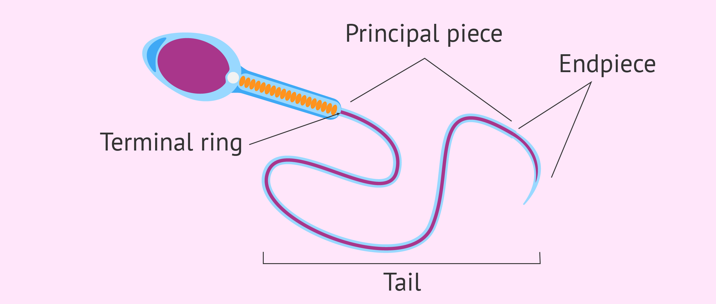
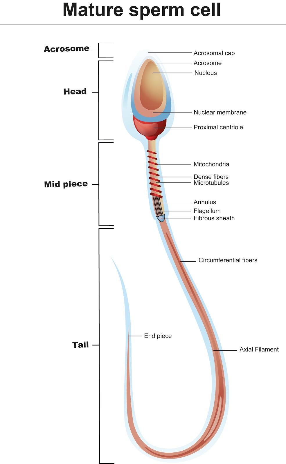
Structure and parts of a sperm cell - inviTRA Structure and parts of a sperm cell 0 This labelled diagram shows the structure of a sperm cellin detail, which has the following parts: Head With its spheric shape, it consists of a large nucleus, which at the same time contains an acrosome. The nucleus contains the genetic information and 23 chromosomes.
Draw a labeled diagram of sperm. - SaralStudy Q:-With a neat diagram explain the 7-celled, 8-nucleate nature of the female gametophyte. Q:-What is oogenesis? Give a brief account of oogenesis. Q:-Describe the structure of a seminiferous tubule. Q:-What is DNA fingerprinting? Mention its application. Q:-What is triple fusion? Where and how does it take place?
An overview of sperm anatomy & morphology | Legacy Sperm is the male sex cell, also known as a gamete. Measuring approximately 0.05 millimeter (0.002 inch) long, sperm cells are made up of a few distinct parts: the tail, made up of protein fibers, which helps it "swim" toward the egg the midpiece, or body, which contains mitochondria to power the sperm's movement
What is a sperm cell like? Its structure, parts and functions - inviTRA Structure and parts of a sperm cell Neck and middle-piece The neck and the middle piece, as the name suggests, are the parts that can be found between the head and the tail. They measure between 6 - 12 microns, a little longer than the head. The width is hardly visible under the microscope. Inside this part are millions of mitochondria.
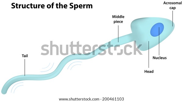
Sperm Cell Labeled Diagram Stock Vector (Royalty Free) 200461103 ... Sperm Cell Labeled Diagram Vector Formats EPS 6733 × 3563 pixels • 22.4 × 11.9 in • DPI 300 • JPG Show more Vector Contributor j joshya Similar images See all Assets from the same collection See all Similar video clips Categories: Science , Food and Drink
Draw a labelled diagram of sperm. - Byju's Solution Sperm: It is the male gamete that is released from the male reproductive organ and is responsible for fertilization in human sexual reproduction. The sperm has a few specific parts Head, a Middle piece, and a Tail. The sperm head contains a very little cytoplasm, an elongated haploid nucleus containing chromosomal material.
Diagram Label Sperm Biology Clip Art - clker.com Search and use 100s of diagram label sperm biology clip arts and images all free! Royalty free, no fees, and download now in the size you need. Facebook Login; X. E-mail Password. ... Animal Cell Labelled. Animal Cell. Cartoon Human Body Parts. First 1 Last. Sub categories to 'diagram label sperm biology'
Male reproductive: The Histology Guide - University of Leeds The production of sperm and eggs/ova (gametes) is a procedure called gametogenesis (spermatogenesis and oogenesis). Gametogenesis involves two rounds of meiotic cell division, in which one diploid cell gives rise to 4 haploid cells.. This diagram shows the processes involved in spermatogenesis. The germinal (seminiferous epithelium) of the seminiferous tubules contains spermatogenic cells and ...
Sperm Under Microscope with Labeled Diagram - AnatomyLearner Sperm Under Microscope with Labeled Diagram 24/06/2022 17/06/2022 by anatomylearner While studying the histological features of the seminiferous tubules and epididymis, you will see sperm cells under the microscope. They are much smaller and lie in groups along the inner margin of the Sertoli cells.
Sperm Diagram Stock Photos, Pictures & Royalty-Free Images - iStock structure of a sperm cell Human Sperm cell Anatomy Penis or Male Reproductive System is a 3D illustration. Comparison between normal and low sperm count Spermatozoons Group Flowing Toward Female Ovum Egg Group of Spermatozoons Flowing Toward Female Ovum Egg Isolated on White Background. Natural Fertilization Process Microscopic View.
The above diagram is of the sperm cell. - Toppr Ask The above diagram is of the sperm cell. (a) Acrosome: It contains enzymes used for penetrating the female egg. ... Draw the diagram of human sperm and label its parts. Write few lines about it. Medium. View solution > View more. More From Chapter. Human Reproduction. View chapter > Revise with Concepts. Advanced Knowledge of Fertilization in ...















Post a Comment for "44 sperm cell diagram with labels"