45 human heart diagram and labels
› heart › picture-of-the-heartHuman Heart (Anatomy): Diagram, Function, Chambers, Location ... Cardiomyopathy: A disease of heart muscle in which the heart is abnormally enlarged, thickened, and/or stiffened. As a result, the heart's ability to pump blood is weakened. As a result, the heart ... › male-human-anatomy-diagramMale Human Anatomy Diagram Pictures, Images and Stock Photos Pacemaker Diagram Cross section of a human heart with pacemaker fitted, showing the major arteries and veins. This is an EPS 10 vector illustration and includes a high resolution JPEG. male human anatomy diagram stock illustrations
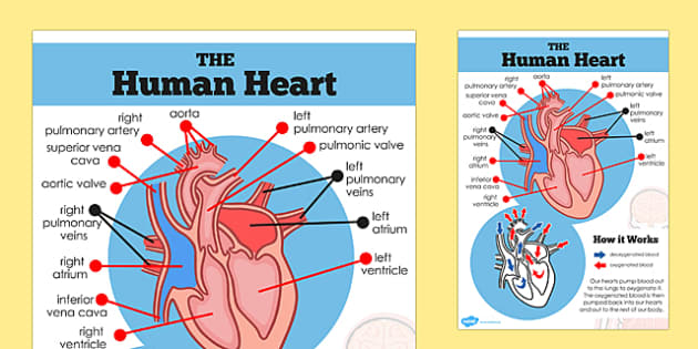
The Human Heart Cardiovascular System Labeling Worksheet - Twinkl This handy heart worksheet gives your children the opportunity to show how much they've learned about this topic. Using the blank heart diagram students are asked to label the aorta, superior vena cava, pulmonary arteries, pulmonary veins, atrium, ventricles, and aortic valves. This simple human heart diagram could be used as both a starter or plenary in order to assess students ...
Human heart diagram and labels
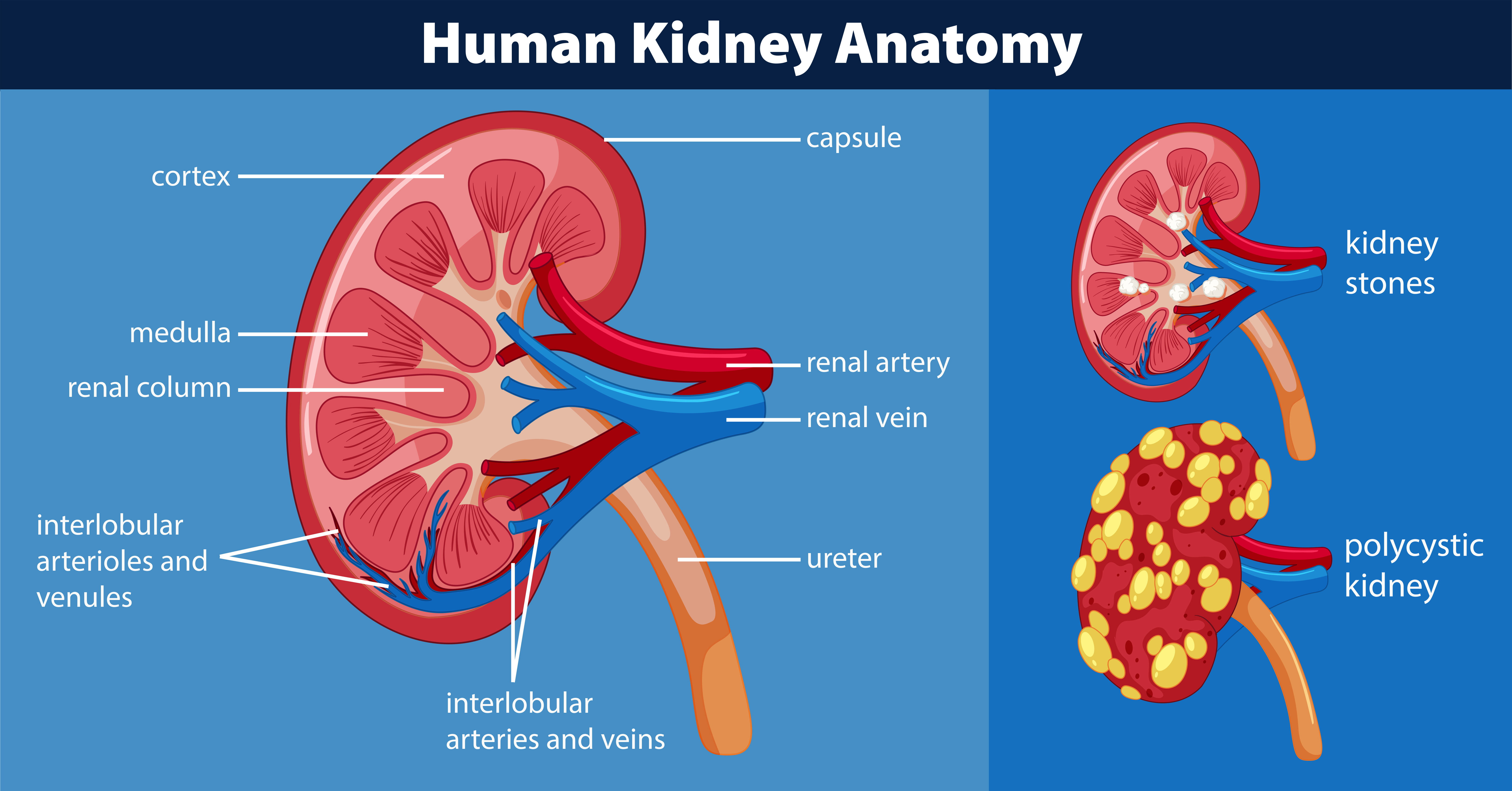
› print › heart-diagramsHeart Diagram – 15+ Free Printable Word, Excel, EPS, PSD ... Teachers and students use the heart diagram, in biological science, to study the structure and functions of a human being’s heart. Friends and colleagues on the other hand may find this diagram template useful when it comes to sending special, personalized gifts to their family members and significant others. Download the template today, and ... cardiovascular system diagram without labels The Cardiovascular System: Anatomy, Physiology, And Adaptations To veteriankey.com. system cardiovascular anatomy exercise blood flow systems body figure physiology adaptations training illustrating main. Solved: Label The Following Diagram Of The Cardiovascular Syste . Label Kidney Diagram - Human Anatomy tartrerepub.blogspot.com byjus.com › biology › diagram-of-heartHeart Diagram with Labels and Detailed Explanation - BYJUS Well-Labelled Diagram of Heart. The heart is made up of four chambers: The upper two chambers of the heart are called auricles. The lower two chambers of the heart are called ventricles. The heart wall is made up of three layers: The outer layer of the heart wall is called epicardium. The middle layer of the heart wall is called myocardium. The inner layer of the heart wall is called endocardium.
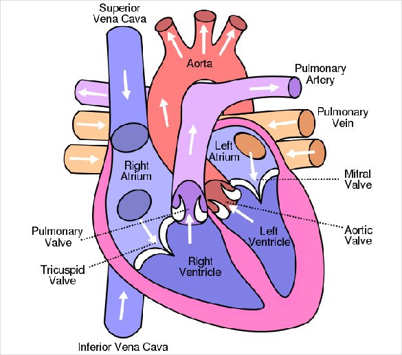
Human heart diagram and labels. en.wikipedia.org › wiki › File:Diagram_of_the_humanFile:Diagram of the human heart (cropped).svg - Wikipedia Add cardiac skeleton. Inferior vena cava more wide. Add aorta in bottom. Add source veins of superior vena cava. Brachiocephalic trunk more wide and separated. Added shadows. Left main pulmonary artery with its first division. 07:02, 2 June 2006: 650 × 650 (26 KB) Yaddah: Diagram of the human heart, created by Wapcaplet in Sodipodi. Cropped by ~~~ to remove white space (this cropping is not the same as Wapcaplet's original crop). Simple heart diagram | Simple heart diagram labeled | Human ... - Pinterest Dec 23, 2021 - Simple heart diagram | Simple heart diagram labeled | Human heart diagram.We provide you a simple heart diagram to draw and learn. Simple heart diagram labeled with accurate labels. Most frequent question in exam to draw human heart diagram with labels. You can learn diagram of heart with labels and easy simple heart anatomy with heart structure. Learn to draw Simple heart ... Human Heart Diagram Pictures, Images and Stock Photos Cross Section of Heart with Labels on White Background Computer generated image of a sagittal cross section view of a human heart, showing chambers, major arteries and veins with anatomy labels. 3d rendering of the human heart anatomy Human heart angioplasty Human heart angioplasty. 3d illustration Anatony of the human heart A Labeled Diagram of the Human Heart You Really Need to See A Labeled Diagram of the Human Heart You Really Need to See Atria and Ventricles. The human heart, comprises four chambers: right atrium, left atrium, right ventricle and left... Valves. The heart features four types of valves which regulate the flow of blood through the heart. These valves have... ...
657 Human Heart Diagram Premium High Res Photos Browse 657 human heart diagram stock photos and images available, or search for heart illustration or pulmonary artery to find more great stock photos and pictures. heart illustration pulmonary artery kidney diagram 11 NEXT File:Heart diagram-en.svg - Wikipedia A labelled diagram of the human heart. Items portrayed in this file ... fix label: 17:12, 14 May 2011: 512 × 403 (277 KB) ZooFari: Add missing Left Atrium label, fix division between aorta and pulmonary arteries. 23:23, 3 August 2010: 470 × 370 (335 KB) ZooFari: Fix septum label: Human Heart Diagram Without Labels - Labelling Worksheet - Twinkl The human heart is a muscle made up of four chambers, these are: Two upper chambers - the left atrium and right atrium Two lower chambers - the left and right ventricles. It's also made up of four valves - these are known as the tricuspid, pulmonary, mitral and aortic valves. How to Draw a Human Heart: 11 Steps (with Pictures) - wikiHow Method 1Sketching the Heart. 1. Draw the lower half of an acorn shape so it's tilted to the left. Use your pen or pencil to start drawing the main part of the heart. This should look like an open-ended acorn that's missing its cap. Draw the shape so it's tilted about 120 degrees to the left.
Label Heart Anatomy Diagram Printout - EnchantedLearning.com EnchantedLearning.comLabel Heart Interior Anatomy Diagram. Human Anatomy. The heart is a fist-sized, muscular organ that pumps blood through the body. Oxygen-poor blood enters the right atrium of the heart (via veins called the inferior vena cava and the superior vena cava). The blood is then pumped into the right ventricle and then through the ... Simple Heart Diagram with Labels Activity - Human Biology - Twinkl The parts of the heart children will learn about in this simple heart diagram with labels are as follows: Aorta. Aortic valve. Left and right atriums. Left and right ventricles. Pulmonary veins. Pulmonary arteries. The above video may be from a third-party source. We accept no responsibility for any videos from third-party sources. The Human Heart Labeling Worksheet (Teacher-Made) - Twinkl The human heart is a muscle made up of four chambers, these are: Two upper chambers - the left atrium and right atrium Two lower chambers - the left and right ventricles. It's also made up of four valves - these are known as the tricuspid, pulmonary, mitral and aortic valves. byjus.com › biology › human-heartHuman Heart - Anatomy, Functions and Facts about Heart - BYJUS The human heart is one of the most important organs responsible for sustaining life. It is a muscular organ with four chambers. The size of the heart is the size of about a clenched fist. The human heart functions throughout a person’s lifespan and is one of the most robust and hardest working muscles in the human body.
Human Heart Labeling Teaching Resources | Teachers Pay Teachers Human Heart Parts and Blood Flow Labeling Worksheets - Diagram/Graphic Organizer by TechCheck Lessons 4.6 (22) $2.25 Zip This resource contains 2 worksheets for students to (1) label the parts of the human heart and (2) Fill in a flowchart tracing the path of blood flowing though the circulatory system. Answer keys included.
Diagram of the human heart royalty-free images - Shutterstock 14,791 diagram of the human heart stock photos, vectors, and illustrations are available royalty-free. See diagram of the human heart stock video clips Image type Orientation Color People Artists Sort by Popular Anatomy Healthcare and Medical Icons and Graphics Diseases, Viruses, and Disorders heart medicine organ hemodynamics circulatory system
Human Heart Diagram Labeled | Science Trends Human Heart Diagram Labeled Anatomy Of The Heart. The human heart usually weighs somewhere between 10 to 12 ounces in men and between 8 to 10 ounces... Pumping Blood Through The Body. The circulatory system surrounding the heart. ... The heart's primary function is to... List Of Heart Structures. ...
commons.wikimedia.org › wiki › File:Diagram_of_theFile:Diagram of the human heart (cropped).svg - Wikimedia Aug 08, 2022 · English: Diagram of the human heart 1. Superior vena cava 2. 4. Mitral valve 5. Aortic valve 6. Left ventricle 7. Right ventricle 8. Left atrium 9. Right atrium 10. Aorta 11. Pulmonary v
Label the heart — Science Learning Hub In this interactive, you can label parts of the human heart. Drag and drop the text labels onto the boxes next to the diagram. Selecting or hovering over a box will highlight each area in the diagram. pulmonary vein. semilunar valve. right ventricle. right atrium. vena cava. left atrium.
A Diagram of the Heart and Its Functioning Explained in Detail The heart blood flow diagram (flowchart) given below will help you to understand the pathway of blood through the heart.Initial five points denotes impure or deoxygenated blood and the last five points denotes pure or oxygenated blood. 1.Different Parts of the Body ↓ 2.Major Veins ↓ 3.Right Atrium ↓ 4.Right Ventricle ↓ 5.Pulmonary Artery ↓ 6.Lungs
Human Heart - Diagram and Anatomy of the Heart - Innerbody The right side of the heart has less myocardium in its walls than the left side because the left side has to pump blood through the entire body while the right side only has to pump to the lungs. Chambers of the Heart. The heart contains 4 chambers: the right atrium, left atrium, right ventricle, and left ventricle. The atria are smaller than the ventricles and have thinner, less muscular walls than the ventricles.
Diagram of Human Heart and Blood Circulation in It Exterior of the Human Heart A heart diagram labeled will provide plenty of information about the structure of your heart, including the wall of your heart. The wall of the heart has three different layers, such as the Myocardium, the Epicardium, and the Endocardium. Here's more about these three layers. Epicardium

Heart Diagrams for Labeling and Coloring, With Reference Chart and Summary | Heart diagram ...
File:Diagram of the human heart (no labels).svg File:Diagram of the human heart (no labels).svg. Size of this PNG preview of this SVG file: 498 × 599 pixels. Other resolutions: 199 × 240 pixels | 399 × 480 pixels | 639 × 768 pixels | 851 × 1,024 pixels | 1,703 × 2,048 pixels | 533 × 641 pixels.
Heart Anatomy: Labeled Diagram, Structures, Blood Flow ... - EZmed There are 4 chambers, labeled 1-4 on the diagram below. To help simplify things, we can convert the heart into a square. We will then divide that square into 4 different boxes which will represent the 4 chambers of the heart. The boxes are numbered to correlate with the labeled chambers on the cartoon diagram.
Human Heart Diagram - Human Body Pictures - Science for Kids Photo description: This is an excellent human heart diagram which uses different colors to show different parts and also labels a number of important heart component such as the aorta, pulmonary artery, pulmonary vein, left atrium, right atrium, left ventricle, right ventricle, inferior vena cava and superior vena cava among others.
Free Unlabelled Diagram Of The Heart, Download Free Unlabelled Diagram Of The Heart png images ...
byjus.com › biology › diagram-of-heartHeart Diagram with Labels and Detailed Explanation - BYJUS Well-Labelled Diagram of Heart. The heart is made up of four chambers: The upper two chambers of the heart are called auricles. The lower two chambers of the heart are called ventricles. The heart wall is made up of three layers: The outer layer of the heart wall is called epicardium. The middle layer of the heart wall is called myocardium. The inner layer of the heart wall is called endocardium.
Human Heart And Their Functions part of the human heart has a very distinct role in blood ...
cardiovascular system diagram without labels The Cardiovascular System: Anatomy, Physiology, And Adaptations To veteriankey.com. system cardiovascular anatomy exercise blood flow systems body figure physiology adaptations training illustrating main. Solved: Label The Following Diagram Of The Cardiovascular Syste . Label Kidney Diagram - Human Anatomy tartrerepub.blogspot.com
› print › heart-diagramsHeart Diagram – 15+ Free Printable Word, Excel, EPS, PSD ... Teachers and students use the heart diagram, in biological science, to study the structure and functions of a human being’s heart. Friends and colleagues on the other hand may find this diagram template useful when it comes to sending special, personalized gifts to their family members and significant others. Download the template today, and ...


Post a Comment for "45 human heart diagram and labels"